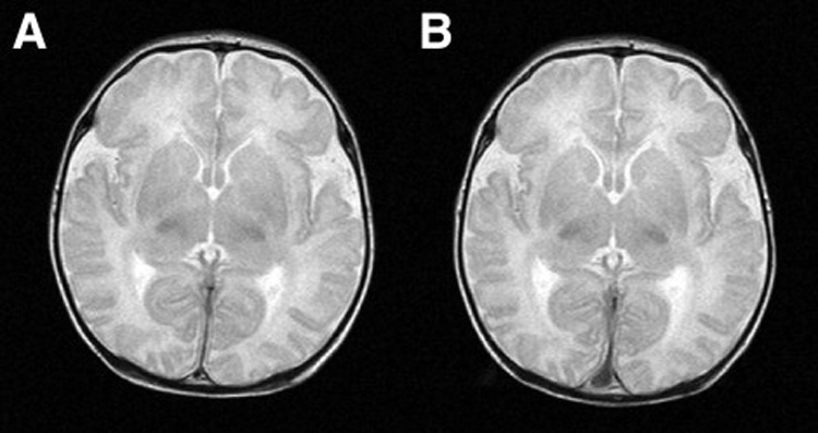
Figure 3.

Figure 3A (left): Normal preoperative T2 weighted sagittal MRI scan in patient with hypoplastic left heart syndrome. Figure 3B (right): The same patient with normal postoperative scan after Norwood Stage I palliation using antegrade cerebral perfusion.
