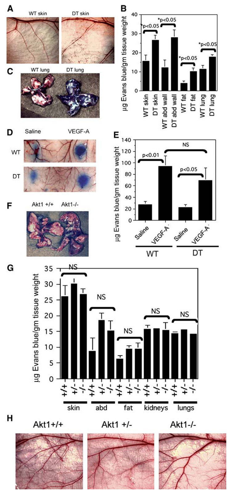
Figure 5. Baseline vascular permeability is increased in double transgenic myrAkt1 mice but not in Akt1 null mice.

A–C: Wild-type (WT) and double transgenic (DT) mice were taken off tetracycline for 5 days prior to analysis of baseline permeability. Evans blue dye (50 mg/kg) was injected i.v. and allowed to circulate for 8 hr, at which time the animals were sacrificed and various organs were collected for analysis. Relatively normal skin vasculature in wild-type and double transgenic mice is shown in A. Evans blue dye was extracted from various organs, and the amount of extracted dye was measured by spectrophotometric absorbance at 620 nm (B). Lungs from WT and DT mice 8 hr after Evans blue injection (C).
D and E: Miles assay in WT and DT mice that were off tetracycline for 5 days. Evans blue dye was injected in the tail vein. Saline or VEGF-A was immediately injected intradermally in the back skin (D). Quantification of Evans blue dye extracted from the skin (E).
F–H: Baseline vascular permeability in Akt1−/− mice and control littermates was determined as described in A. F: Whole lungs taken from Akt1+/+ and Akt1−/− mice 8 hr after Evans blue injection. G: Quantitation of extravasated Evans blue in various organs from Akt1+/+, Akt1+/−, and Akt1−/− mice. H: Normal vasculature is seen in the flank skin in these mice. Data from all the experiments above represented four mice per group and were calculated as micrograms of dye per gram tissue weight (mean ± SEM). p value < 0.05 was considered statistically significant. NS is not statistically significant.
