
Figure 9.
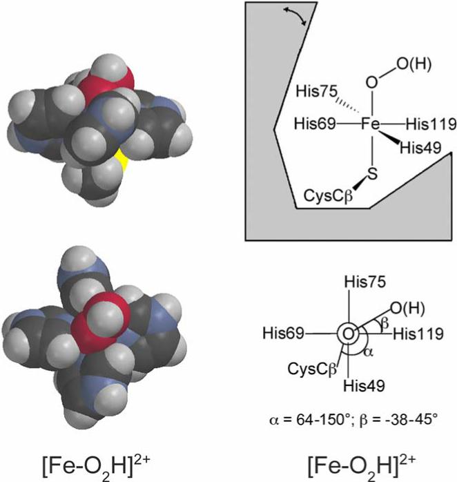
Space-filling depictions of DFT-optimized modeled structures of the putative ferric-(hydro)peroxo intermediate (left), and schematic diagrams showing the geometric restrictions of this intermediate at the SOR site (right), modified with permission from Ref. (5). The modeled structures used imidazoles and methyl thiolate in place of His and Cys ligands, respectively, and the torsion angles of the imidazole rings around the Fe-N bonds were fixed at those in the D. desulfuricans 2Fe-SOR crystal structure (PDB ID 1DFX)(18). Color coding of atoms is carbon, black; hydrogen, gray; oxygen, red; nitrogen, blue; sulfur, yellow, iron, orange. The double-headed arrow indicates movement of the flexible loop region.