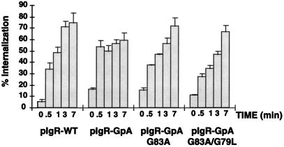
Figure 5.
Receptor-mediated internalization of [125I]dIgA. MDCK cells expressing pIgR-WT or the chimeras were grown on permeable supports for 4 d. Cells were incubated at 4°C with [125I]dIgA at the basal surface for 1 h, washed, placed at 37°C for the given times, and then rapidly cooled back to 4°C. The cell surface was then stripped of remaining dIgA and the filters were counted. Calculations were based on the percent IgA internalized/total dIgA bound at time zero (stripped from the cell surface after internalization + dissociated into the basal media during warming at 37°C + internalized). Single clones were used for this experiment (n = 6), error bars represent the SD.

