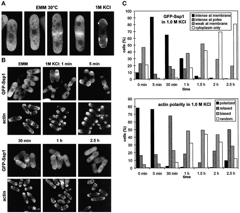
Figure 3.
Cellular localization of GFP-SspI. (A) Strain Q1591 (wild type harboring the pIR2–22 plasmid) was grown in EMM lacking thiamine (EMM, 30°C) and then transferred to EMM + 1 M KCl. A sample was collected from this culture after 10 min of incubation at 30°C. Cells were analyzed by confocal microscopy. (B) The same strain was grown in EMM as in A and then shifted to EMM + 1 M KCl and incubated at 30°C. Cells were taken at the indicated times, observed to localize GFP-Ssp1, and also fixed and stained with TRITC–phalloidin to visualize filamentous actin. Images were taken using the fluorescence microscope. (C) Quantitative scoring of the cell population in experiment B. The presence of GFP-Ssp1 at the plasma membrane (top graph) was scored in living cells; the polarity of actin patches (bottom graph) was blind-scored in cells fixed and stained for actin (n ≥ 200). Intense at membrane: distinct rim of fluorescence along the surface. intense at poles: distinct only at poles, none or threshold of detection in the middle; weak at membrane: weak rim all over the surface or at poles only; polarized: wild-type aggregations of actin patches at cell ends; relaxed: relaxed aggregations, more patches outside poles; biased: patches all over the cell surface but bias toward the poles maintained; random: completely random distribution.

