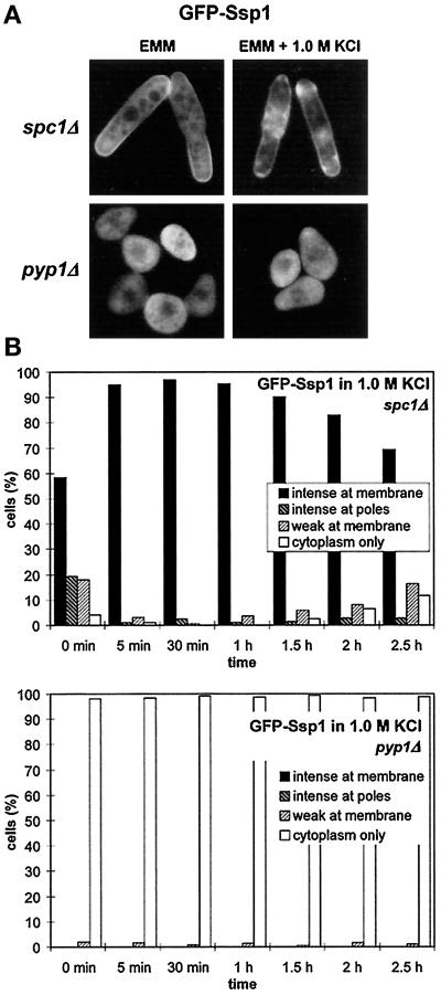
Figure 7.
Ssp1 localization depends on the activity of the Spc1 MAP kinase. (A) Ssp1 shows increased localization at the plasma membrane in the spc1Δ background and reduced localization in the pyp1Δ background. Strains Q1580 (spc1Δ) and Q1592 (pyp1Δ), both harboring the pIR2–22 plasmid, were grown overnight at 30°C in EMM lacking thiamine. Half of each culture was then transferred to the presence of 1 M KCl in the same medium and incubated for another 10 min. All four samples were then taken, and the GFP-Ssp1 fluorescence was observed. (B) A time course of the GFP-Ssp1 redistribution after transfer to 1 M KCl in experiment A. For explanation of categories and wild-type control, see Figure 3C (n ≥ 200).

