Figure 5.
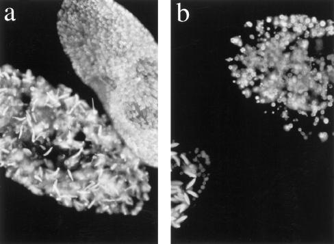
Phenotypic characterization of cells transformed with T1 and T4 coding sequences. Immunofluorescent images of trichocysts of a pT1b-transformed cell beneath an uninjected wild-type cell (a) and pT4a-transformed cells (20–25 divisions after microinjection) (b). Both pT1b- and pT4a-transformed cells presented undocked aberrantly shaped trichocysts, which tended to swell more readily than wild-type trichocysts under the fixation conditions used. Magnification, 500×.

