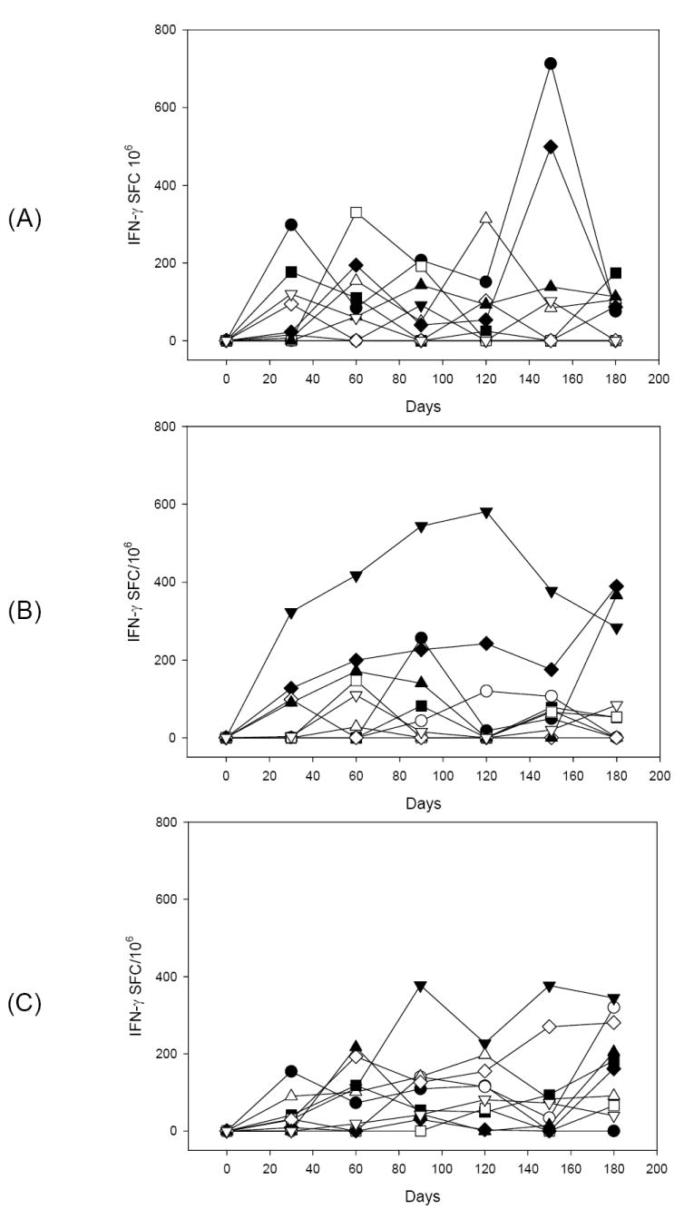
Figure 3.

Kinetics of the prevalence of IFN-γ secreting cells determined by ELISPOT in PBMC obtained from animals immunized with different formulations of rPvRII after ex vivo stimulation with the recombinant protein. Data are presented for individual animal at different time points and expressed as IFN-γ spot forming cells per 1×10⁶ PBMC. Average values for medium control wells in the presence of cytokines were initially subtracted from the average values obtained from antigen-stimulated wells. To take in consideration individual variability, recall PvRII-reactive T cells were calculated by subtracting the spot forming cells obtained with pre-immune samples. Rhesus macaques were immunized with 50 μg P. vivax DBP RII (closed symbols) or 10 μg P. vivax DBP RII (open symbols) formulated with Alhydrogel (A), Montanide ISA 720 (B) or AS02A (C).
