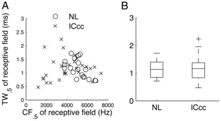
FIG. 6.

Temporal widths (TWs) of the receptive fields. A: TW0.5 of the STRFs of NL (circles) and ICcc neurons (crosses) plotted against the CF0.5. There was no apparent correlation between the parameters for ICcc and a weak and possibly artifactual correlation for NL (see text). B: boxplot of the distribution of temporal widths seen in the two nuclei. Median TW0.5 was not significantly different (P > 0.7).
