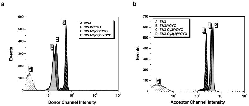
Figure 8.
Flow cytometry results recorded for 4 different bead populations monitoring the donor channel (a) and acceptor channel (b). Beads labeled with 3WJs but no intercalating dye gave only background signal (A). Beads labeled with 3WJs and YOYO-1 (B) gave strong signals in both channels due to the broad emission spectrum of the intercalator. Addition of one (C) or two (D) Cy3 acceptor dyes led to a decrease in intensity in the donor channel due to efficient FRET (left). A corresponding increase in fluorescence was observed in the acceptor channel due to sensitized emission from the Cy3 labels (right).

