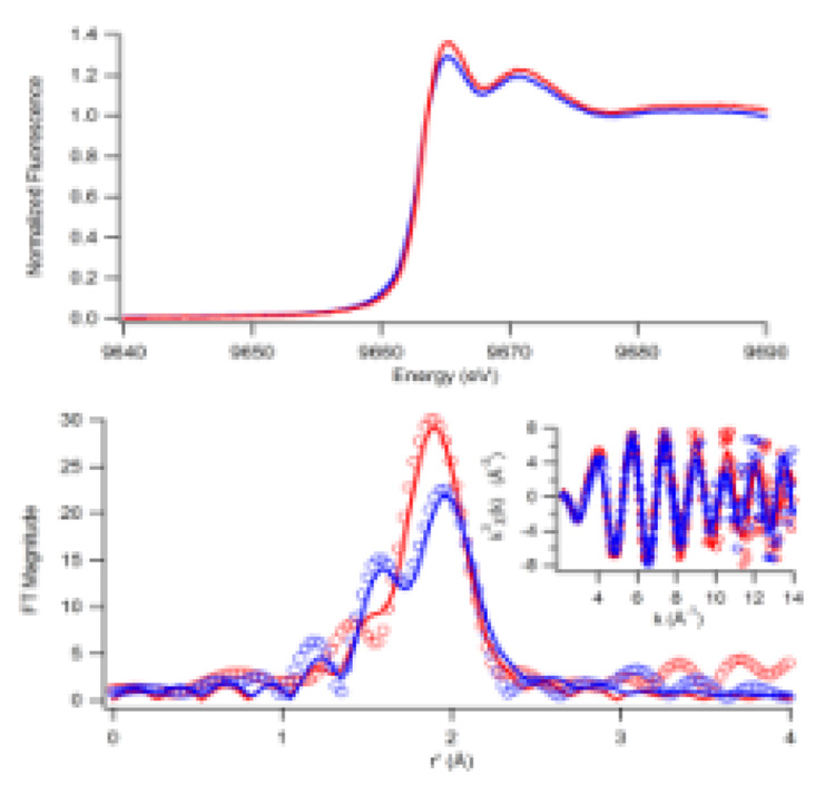
Figure 1.
Zn K-edge XAS of HpHypA without a second metal (blue) and with Ni(II) (red). Top: The XANES spectra show a small shift in the relative intensities of the first two peaks. Bottom: The Fourier-transformed EXAFS spectra (FT window = 2–14 Å−1, uncorrected for phase shifts, bottom) show that in the Ni-free sample a second peak corresponding to an N/O-donor is present (data shown as circles), unfiltered exafs data is shown in the inset. Fits calculated for the apo-protein 1N/O @ 2.03(3) Å (σ2 = 0.002(2) Å2) + 3S @ 2.34(1) Å (σ2 = 0.003(1) Å2), GOF = 0.88, and for the protein with Ni added, 4S @ 2.32(1) Å (σ2 = 0.003(1) Å2), GOF = 0.78, are shown as solid lines (see supporting information).

