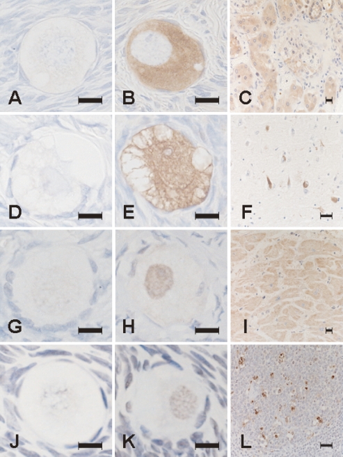
Fig. 1.
Photomicrographs of sections processed with immunohistochemical staining for pentosidine (A–C), ubiquitin (D–F), caspase 12 (G–I) and TUNEL stainings (J–L). Positive signals for these substances were undetectable on negative reaction control sections for pentosidine (A), ubiquitin (D), caspase 12 (G), and TUNEL (J). Pentosidine immunoreactivity is localized in the cytoplasm of oocytes (B) and in the cytoplasm of epithelial cells of proximal tubuli in the kidney of diabetic nephropathy as a positive reaction control (C). Ubiquitin immunoreactivity is localized in the cytoplasm and nucleus of oocytes (E) and in neurofibrillary tangles in the cerebral cortex of Alzheimer’s disease as a positive reaction control (F). Caspase 12 immunoreactivity is localized in the nucleus of oocytes indicative of nuclear translocation of the activated form (H) and in the cytoplasm of cardiocytes of the heart as a positive reaction control with a dotlike pattern indicative of microsomal localization of the inactive form (I). TUNEL positivity is localized in the nucleus of oocytes (K) and in large lymphocytes in the germinal center of a mucosal lymph follicle in catarrhal appendicitis as a positive reaction control (L). Bar=50 µm.

