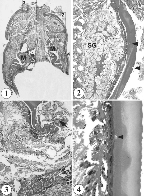
Fig. 1-4.
Histological sections of the tick extracted from the scalp of a Korean boy. 1. A whole section of the tick (up; capitulum side, down; tail side) showing its characteristic body contour, cuticle structures, and well developed musculature (M). H-E stain, × 40. 2. An enlarged view of box 2 in Fig. 1 showing well developed salivary glands (SG) and cuticle with pore canals (arrowheads). H-E stain, × 100. 3. An enlarged view of box 3 in Fig. 1, the capitulum side. A part of the capitulum (arrow) is seen sectioned. H-E stain, × 100. 4. An enlarged view of box 4 in Fig. 1 showing the characteristic saw-like cuticular surface and a pore canal. H-E stain, × 400.

