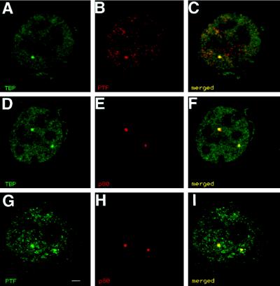
Figure 2.
Optical sections of immunofluorescently double-labeled T24 cells are shown. (A and D) Labeling of TBP shows the transcription factor to be distributed throughout the nucleus and concentrated in a few small nuclear domains. (B and G) Labeling for PTFγ gives a similar overall distribution and is concentrated in a few nuclear foci. (C) When the labeling patterns are compared, the brightly labeled domains colocalize. (E and H) Labeling for p80-coilin reveals coiled bodies. When the p80-coilin labeling is compared with the TBP labeling (F) or the PTFγ labeling (I), the transcription factor-enriched domains colocalize with coiled bodies. Bar, 2 μm.

