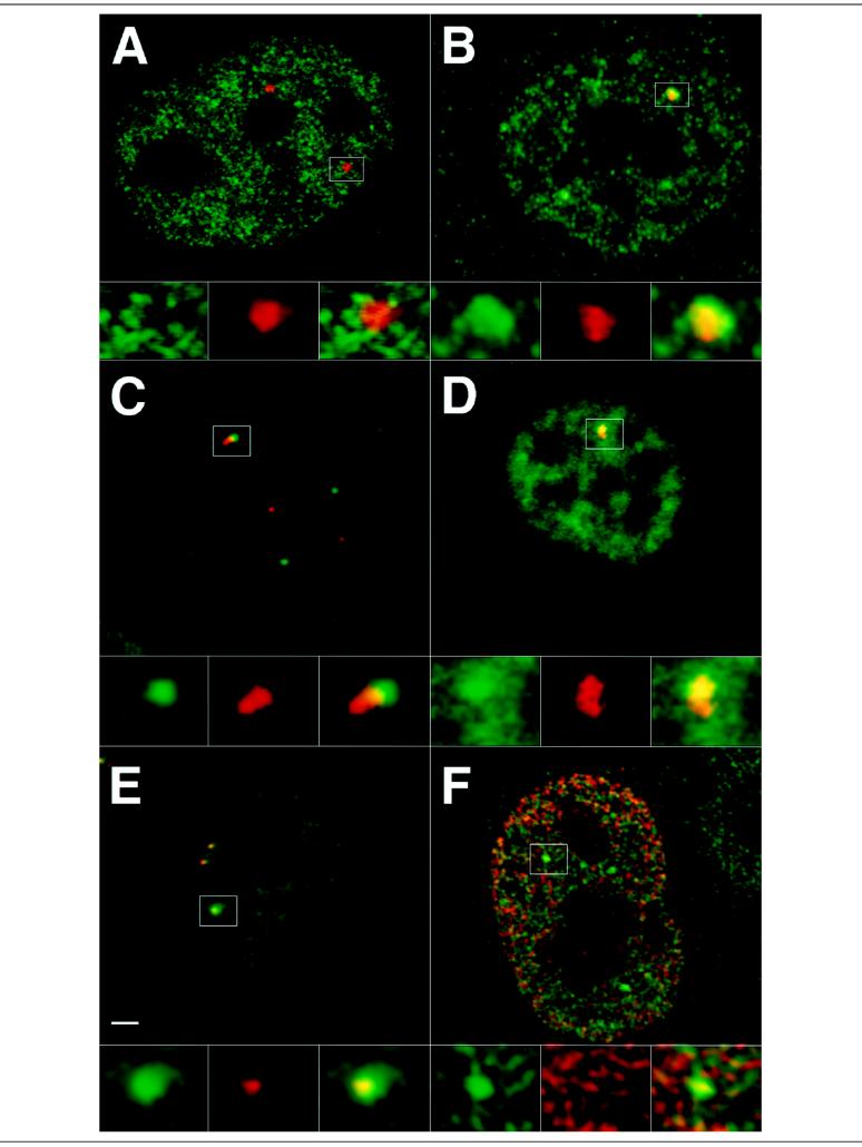
Figure 4.
Optical sections of immunofluorescently double-labeled cells are shown. (A) Labeling with the H5 antibody against the hyperphosphorylated form of RNA polymerase II (green) reveals a punctated pattern throughout the nucleus. A combined p80-coilin labeling of coiled bodies (red) shows that the H5 dots do not overlap with the coiled body, although a number of dots can be found at the periphery of the coiled body (compare separate channels in magnified area). (B) Labeling with 8WG16 antibody against the hypophosphorylated form of RNA polymerase II (green) also reveals a punctated pattern in addition to a few brightly labeled domains. Double labeling with p80-coilin shows that these domains are closely associated with coiled bodies, similar to the transcription factor-enriched domains (see Figure 3) (compare separate channels in magnified area). (C) Immunofluorescent labeling for p80-coilin (green) was combined with genomic in situ hybridization, visualizing the U2 snRNA gene loci (red). A coiled body could be found adjacent to a U2 snRNA gene locus in about 30% of the cases. (D) Brightly labeled 8WG16 domains (green) could be found colocalizing with U2 snRNA gene loci (red). (E) Also domains enriched in the transcription factor PTFγ (green) were found overlapping with some U2 snRNA gene loci (red). (F) Newly synthesized RNA was visualized by microinjecting cells with BrUTP (red). Double labeling with 8WG16 (green) shows that there is active transcription inside the domain enriched in hypophosphorylated RNA polymerase II (compare separate channels in magnified area). Bar, 2 μm.

