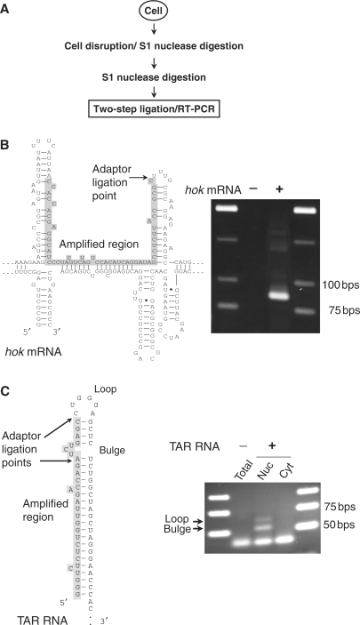
Figure 5.
Mapping of dsRNA regions in cellular RNA. (A) Experimental flowchart of extraction and detection of natural dsRNA fragments from cells. Fragments of dsRNA were ligated to Y-adaptors and subjected to RT–PCR amplification. (B) Detection of a dsRNA/ssRNA junction at the loop within folded hok mRNA (PCR product is 87 bp). (C) Detection of dsRNA/ssRNA junctions at the internal bulge and the loop within the TAR RNA element (PCR products are 55 and 62 bp, respectively) in RNA from nuclear (Nuc) and cytoplasmic (Cyt) fractions. Structures of hok and TAR RNAs, the amplified dsRNA region and adaptor ligation points are indicated.

