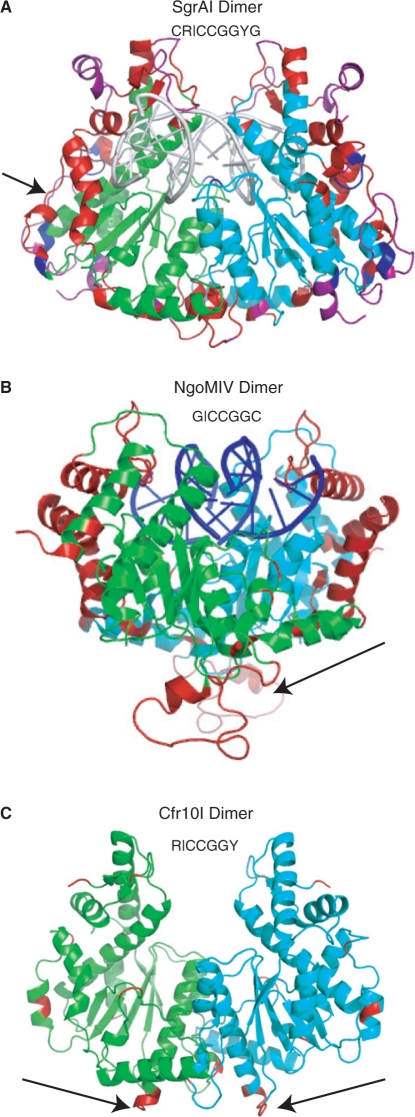
Figure 1.
Dimer structures of SgrAI, NgoMIV and Cfr10I. (A) SgrAI dimer with two subunits in green and cyan. Regions that do not align with NgoMIV, Cfr10I or either, are shown in red, blue and purple, respectively. Arrow indicates the segment corresponding to the loops of NgoMIV that form the dimer–dimer interface. DNA shown as a cartoon in white. (B) NgoMIV dimer with two subunits in green and cyan. Regions that do not align with SgrAI are shown in red. The arrow indicates loops that form the dimer–dimer interface. Bound DNA is shown as a cartoon in blue. (C) Cfr10I dimer with two subunits in green and cyan. Regions that do not align with SgrAI are shown in red. The arrows indicate the extensions of the alpha helices involved in dimer–dimer contacts.

