Figure 7.
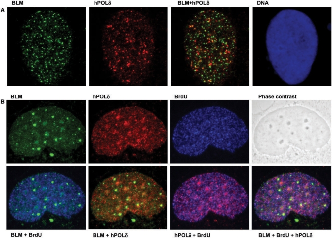
Dual staining for BLM and hPOL δ suggests co-localization in vivo, which is stimulated during replicative stress. (A) GM00637 cells were arrested with 2.5 mM HU, and were stained as described in Materials and methods section. A representative image showing punctate BLM (green, left) and hPOL δ (red, second from left) staining. Coincidence of the green and red signals (yellow signal) suggests co-localization of the two proteins. The right panel shows the nucleus of the same cell stained with the DNA dye Hoechst 33258. (B) GM00637 cells grown on coverslips without HU treatment and were pulse-labeled with 25 μM BrdU for 5 min, and then stained as described in Materials and methods section. Representative images show staining of the same nucleus for BLM in green (top left), for hPOL δ in red (top, second from left), for BrdU in blue (top, second from right). Phase contrast image of the same nucleus is depicted on the top right. In the bottom row, combined images of the individual stainings from the top row are shown. As expected, the BrdU signal correlates well with the signal for hPOLδ (magenta-color pattern second panel from bottom right). The coincidence of BLM signal with either BrdU (cyan signal in bottom left) or hPOLδ (yellow signal in second panel from bottom left) is less pronounced.

