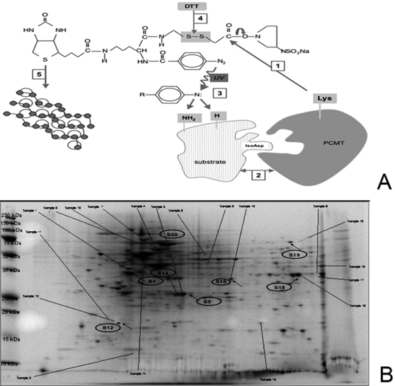
Figure 5. Identification of PCMT substrates.
Panel A: Experimental strategy for isolation and characterization of PCMT substrates. Step 1: human recombinant PCMT, purified to homogeneity, was immobilized onto sulfoSBED by N-hidroxysuccinimide chemistry; Step 2: cell extracts as a source of substrates were added and Step 3: proteins interacting with PCMT were immobilized upon UV photoactivation; Step 4: PCMT was released and biotin “transferred” onto the methyltrasferase substrate (Label Transfer Method); Step 5: purification was accomplished by exploiting streptavidin-biotin interactions. Panel B: 2D gel electrophoresis imaging of comparative proteomics. HUVEC were infected with antisense PCMT carrying retrovirus and then stressed with 0.3 mM of H2O2. Cells lysates were reacted with Sulfo-SBED previously cross-linked with recombinant PCMT (Panel B). Arrows indicate the protein spots which have been characterized as reported in Table 1. Background noise due to aspecific binding was subtracted by comparison with the 2D image obtained from a parallel sample reacted with non-cross-linked Sulfo-SBED.

