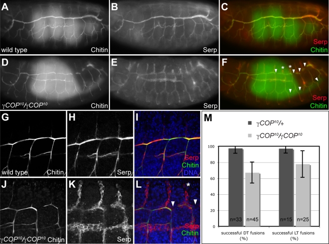
Figure 6. γCOP is required for apical protein secretion and tracheal lumen morphogenesis.
(A–F): Chitin and Serp protein accumulate in the tracheal lumen of stage 16 wild type embryos (A–C). In contrast, Serp protein is retained in tracheal cells in γCOP10 mutants, while luminal accumulation of chitin is not affected in the mutants (D–F). Note that the tracheal lumen marked by chitin staining in γCOP10 embryos (D) is narrower than in wild type embryos (A). In addition, γCOP10 embryos display interruptions in the DT lumen and in the LT (arrowheads in F) and stunted dorsal branches (asterisks in F) in posterior metameres. Also note that γCOP10 embryos are developmentally delayed compared to wild type embryos, as indicated by gut morphology (green gut autofluorescence is visible in C, F). (G–L): Close-ups of wild type (G–I) and γCOP10 (J–L) embryos. DT lumen fusion defects (arrowheads) and DB migration defects (asterisk) are indicated in (L). (M): Quantification of DT and LT fusion defects in γCOP10 embryos (light grey bars) compared to heterozygous siblings (dark grey bars). 100% corresponds to nine successful fusion events in the ten tracheal metameres on one side of the embryo. Error bars indicate standard deviation. (A–F) are wide field fluorescent micrographs, (G–L) are single confocal sections taken at identical settings.

