Figure 2.
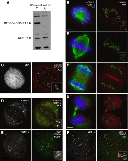
CENP-V localizes to kinetochores throughout mitosis. (A) Immunoblot of HeLa cell lysate probed with serum Ra4552. T, lysate from HeLa cells expressing CENP-V–GFP–TrAP; U, lysate from untransfected HeLa cells. (B–B′′′) Localization of endogenous CENP-V (red) to kinetochores in (B, B′) prometaphase and metaphase, (B′′) to the mid-zone in anaphase and (B′′′) the mid-body in cytokinesis in HeLa cells (microtubules, green; DNA, blue). Right panels show CENP-V (red) co-stained with anticentromere antibody (ACA, green). (C) Localization of endogenous CENP-V (green) to kinetochores co-stained with Aurora B (red) and ACA (white), in cells treated with colcemid (2 h); (D–F) Localization of endogenous CENP-V (green (D, E), red (F)) co-stained with various centromere/kinetochore markers as follows: (D) CENP-A (red), (E) Hec1 (red), INCENP (white), (F) CENP-E (green) and the plus end MAP EB1 (white). Insets show higher magnification views of a single optical section of the same region. Bars, 5 μm.

