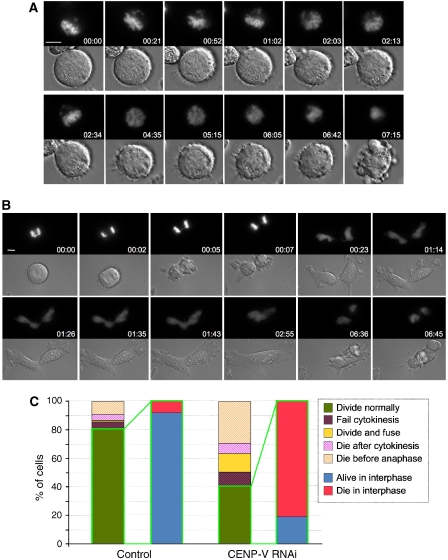
Figure 5.
Live imaging of CENP-V-depleted cells reveals defects in chromosome alignment and cytokinesis. (A, B) Selected fluorescence and DIC frames of live cell imaging experiments performed on a mitotic HeLa cell line stably expressing histone H2B–GFP treated with a specific CENP-V siRNA oligonucleotide. (A) Some cells are incapable of establishing a correct alignment in metaphase and fail to enter anaphase. (B) Other cells manage to enter anaphase with chromatin bridges and fail cytokinesis. (C) Quantification of the various phenotypes observed when CENP-V is depleted. Bar, 5 μm.

