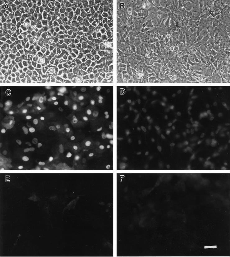
Figure 1.
Micrographs of primary cultures of 14-d corneal epithelial cells (A and C) and corneal fibroblasts (B and D), and 8-d skin epithelial cells (E) and skin fibroblasts (F). Panels A and B are phase contrast. Panels C–F are immunofluorescence micrographs of cells reacted with the anti-chick ferritin monoclonal antibody 6D11. Bar, 25 μm.

