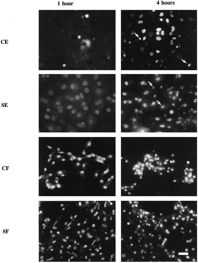
Figure 3.
Fluorescence micrographs of 12-d corneal epithelial cells (CE), 8-d skin epithelial cells (SE), 12-d corneal fibroblasts (CF), and 8-d skin fibroblasts (SF) reacted for DNA breaks by ISEL. The cells were exposed to UV light for 5 min and then were fixed at 1 h and 4 h after treatment. Arrows indicate the labeling of small nuclei or nuclear fragmentation. Bar, 25 μm.

