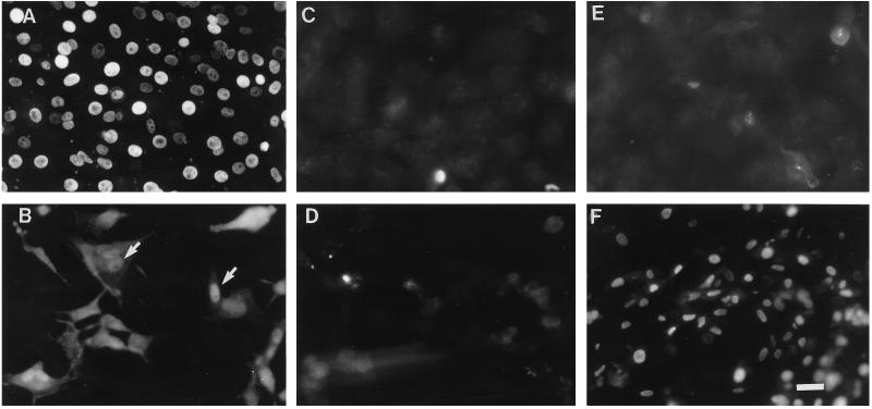
Figure 6.
Fluorescence micrographs of 12-d corneal epithelial cells (A, C, and E) and corneal fibroblasts (B, D, and F) cultured in normal (C and D) or high-iron medium (100 μM ferrous sulfate) (A, B, E, and F) for 16 h before a 3-min UV exposure. The cells were processed 4 h later for either ferritin localization or DNA breaks by ISEL. Panels A and B are the immunofluorescence micrographs of cells reacted for ferritin with monoclonal antibody 6D11. In the fibroblasts, both nuclear (arrows in panel B) and cytoplasmic immunoreactivity was observed. Panels C, D, E, and F are the micrographs of cells examined for DNA breakage by ISEL. Bar, 25 μm.

