Figure 7.
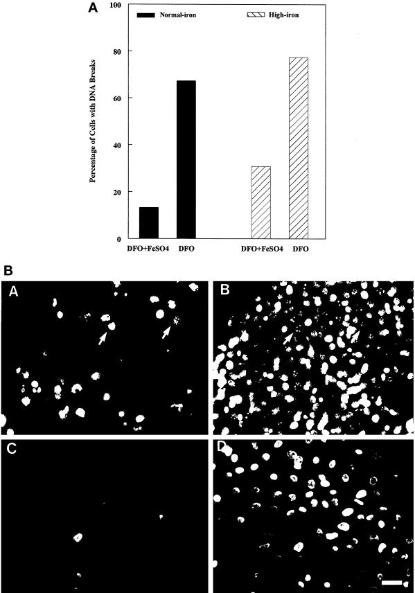
(A) (top panel), Quantitation of UV-induced DNA breaks in day 7–8 corneal epithelial cells treated with 100 μM deferoxamine (DFO) or equimolar concentrations of deferoxamine and ferrous sulfate (DFO+FeSO4). Both groups of cells were cultured for an additional 16 h in normal-iron medium (solid bars) or high-iron medium (hatched bars). They were then exposed to UV light for 5 min and fixed 4 h later. The quantitative data were derived by averaging the percentage of positive cells for DNA breaks from two separate experiments. (B) (bottom panel), Fluorescence micrographs of 8-d corneal epithelial cells examined for DNA breakage by ISEL (A and C), and by Hoechst staining (B and D). The cells were treated either with deferoxamine (100 μM) (A and B) or equimolar concentrations of deferoxamine and ferrous sulfate (C and D) for 48 h. They were then incubated in normal-iron medium for an additional 16 h before exposure to 5 min of UV irradiation and fixation 4 h later. In the deferoxamine-treated cells, stronger ISEL signals and more nuclear fragmentation were observed (A, arrows), as also seen in Hoechst staining (B, arrows). Bar, 25 μm.

