
Figure 7.
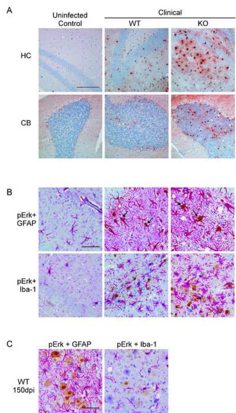
Determination of cells expressing p-Erk1/2 in the brains of scrapie-infected mice. (A) Shown are representative sections of the hippocampus (HC) and cerebellum (CB) from mock-infected controls and scrapie-infected clinical CCR1 KO mice stained for p-Erk1/2 expression. Magnification bar represents 200µm. (B) Sections were then co-stained with either GFAP or Iba-1, which stains the cytoplasms of activated astrocytes or microglia, respectively (bright pink around the nuclei). Cells expressing p-Erk1/2 are stained with DAB, (shown in brown). All sections were counterstained with hematoxylin. Arrows indicate colocalization of p-Erk1/2 expression with the anti-GFAP stain. This co-localization was not seen with anti-Iba-1. Magnification bar represents 100µm. (C) Dual staining of scrapie-infected WT cortex at 150dpi showing p-Erk1/2 staining in large neuron-like cells which were not co-stained by anti-GFAP or anti-Iba-1. Magnification bar represent 100µm.