
Figure 8.
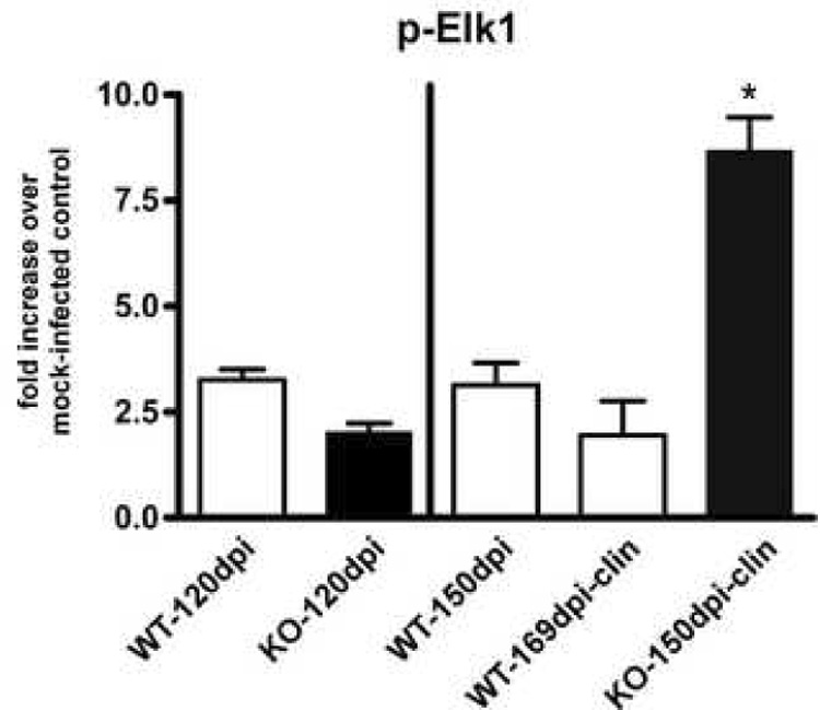
Quantitation of p-Elk-1 in scrapie-infected CCR1 KO and WT mice. Brains from scrapie-infected mice were analyzed for amounts of phosphorylated Elk-1 protein by Western blot and quantitated by enhanced chemifluorescence. Data is expressed as fold increase over mock-infected controls. p-Elk-1 is significantly increased in the clinical CCR1KO mice as compared to WT controls at 150 dpi as well as at the clinical time point (Mann-Whitney test; p<0.05).