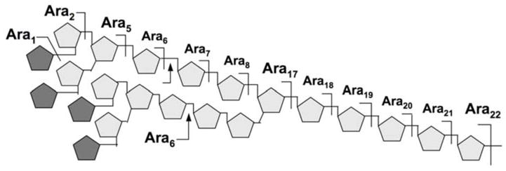
Fig. 1.

The Ara22mer structural model for AG. The proposed branching pattern was based primarily on the absence of Ara3,4 and Ara9-16 among the single chemical cleavage fragments detected by MS, as indicated above the 5-arabinan chain. Apart from the well established non-reducing terminal Ara6 motif, which can be released intact by the Cellulomonas arabinananse (indicated by arrows), the overall assembly of the arabinan motif remains speculative. The pentagons represent α-Araf residues, with darker shaded ones representing β-Araf.
