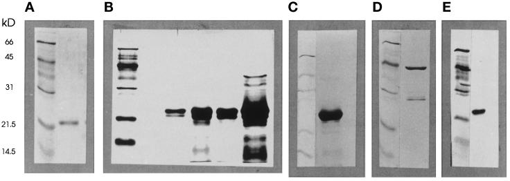
Figure 1.
Analysis of recombinant GTPases. All recombinant proteins used in this work were analyzed for purity by electrophoresis in 12% SDS-polyacrylamide gels and detection by silver staining. The panels illustrate the positions of Mr markers as indicated together with (a) Rac2; (b) Cdc42/G25K (four preparations); (c) N-Myc-Cdc42/G25K; (d) N17-Rac2-GST with GST; (e) N17-Cdc42. Note the heterogeneity of Cdc42/G25K, variable between preparations (b) and the relative homogeneity of the N-Myc tagged Cdc42/G25K product (c).

