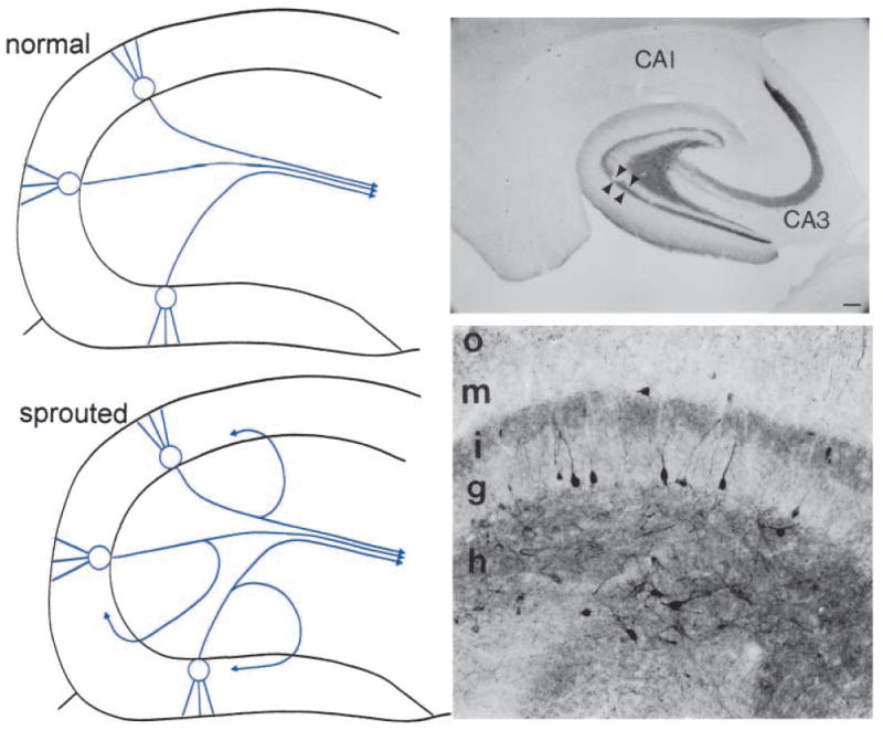
Fig. 1.

Mossy fiber sprouting. Left: Top. A schematic of the normal dentate gyrus granule cell projection, the mossy fibers, which mainly target CA3 pyramidal cells. Not shown are collaterals that innervate hilar dendrites of nonprincipal cells (interneurons and mossy cells of the dentate gyrus). Bottom. A schematic of the dentate gyrus granule cell axons after seizures that induce mossy fiber sprouting. Collaterals of the mossy fiber axons sprout into the inner molecular layer, where they are thought to innervate proximal dendrites of granule cells and inhibitory neuronal processes. Right: A, Mossy fiber sprouting from a pilocarpine-treated rat with recurrent seizures, illustrated by Timm stain for heavy metals. The sprouted fibers are stained darkly in the inner molecular layer (IML). Mossy fibers are also stained in the hilus (HIL). GCL = granule cell layer. Calibration = 100 μm. B, A section from a pilocarpine-treated rat that had status epilepticus and recurrent seizures, that was stained using an antibody to neuropeptide Y. Sprouted fibers are indicated by the arrows. DG = dentate gyrus. Calibration (in A) = 250 μm.
