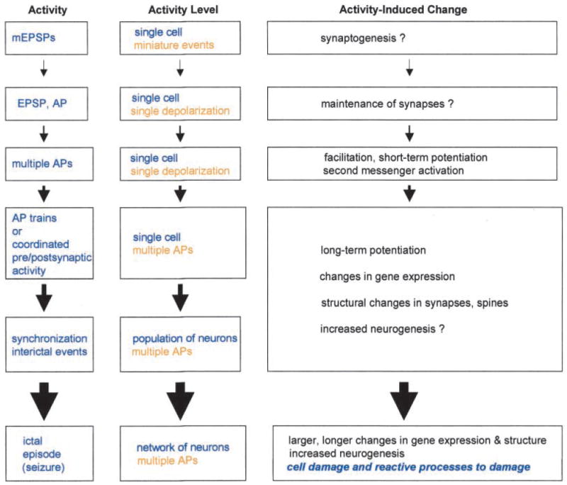
Fig. 4.

Activity-dependent changes in the dentate gyrus as a continuum. A diagram illustrates the concept of a continuum from plasticity induced by simple forms of neural activity, such as a single depolarization in a single cell, to the changes following seizures. Physiological levels of activity are listed from top to bottom, in increasing order. The actual physiological events are described according to the number of neurons involved and extent of depolarization in the center column, as the “Activity level.” Examples of the types of plastic changes associated with each level of activity are listed on the right as “Activity-induced changes,” some of which are not completely clear (“?”). Set apart from the rest is the one major difference between seizures and lesser forms of activity, at least as currently understood, which are the changes that follow cell death or damage due to seizures (see text). EPSP = excitatory postsynaptic potential; mEPSP = miniature EPSP; AP = action potential.
