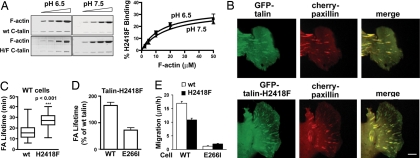
Fig. 5.
Talin H2418F has reduced pH-dependent affinity for actin binding and alters FA duration and migration rate. (A) Cosedimentation of 1 μM WT USH-I/LWEQ or USH-I/LWEQ containing an H2418F mutation with increasing actin concentrations after incubation for 60 min at pH 6.5 or pH 7.5. Shown are Coomassie-stained gels of pellet fractions (Left) and kinetics of talin binding as a function of F-actin concentration (Right). (B) Images from movies of WT cells showing colocalization of cherry-paxillin and GFP-talin or GFP-talin-H2418F at the edge of a wounded monolayer 6 h after wounding. (Scale bar: 5 μm.) (C) Lifetime of FAs in WT cells was determined by analysis of cherry-paxillin in 10–15 FAs in movies from five independent cell preparations. (D) Lifetime of FAs in WT and E266I cells expressing full-length talin-H2418F relative to expression of WT talin in cells at the edge of a wounded monolayer. (E) Migration rate of wound-edge WT and E266I cells expressing GFP-talin or GFP-talin-H2418F determined as distance traveled for 10 h after wounding by tracking individual cells. Data are expressed as means ± SEM of transfected cells in movies from three independent cell preparations.

