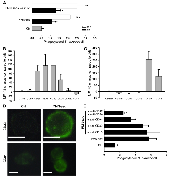
Figure 2. PMN-sec enhances activation of macrophages and expression of FcγRs.
(A) Human macrophages were incubated with PMN-sec for either 2 or 24 hours or with medium. In some wells, the PMN-sec was washed off after the 2- or 24-hour incubation period and replaced by medium. Subsequently the phagocytic activity was quantified. For each analysis, 4 independent experiments were performed. *P < 0.05 versus control; **P < 0.05 versus control and the respective 2-hour treatment group. (B and C) Expression of activation markers (B) and phagocytic receptors (C) in macrophages in response to treatment with PMN-sec for 24 hours. Expression is given as percentage change compared with basal expression. All values are isotype corrected. For each analysis, 6–8 independent experiments were performed. (D) Representative images of antibody staining for CD32 and CD64 in macrophages after treatment with PMN-sec or medium. Scale bar: 10 μm. (E) Human macrophages were incubated with PMN-sec or medium for 24 hours. Blocking antibodies toward CD64, CD32, or CD16 were added 30 minutes before incubation with IgG-opsonized S. aureus. The number of phagocytosed bacteria after 1 hour was quantified. For each analysis, 6 independent experiments were performed. †P < 0.05 versus group treated with PMN-sec.

