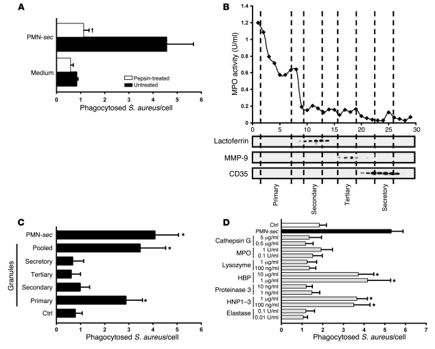
Figure 3. Identification of active PMN granule components.
(A) PMN-sec was digested with pepsin and the remaining activity was tested. For each analysis, 4 independent experiments were performed. †P < 0.05 versus group treated with active PMN-sec. (B) Localization of neutrophil granules in fractions (x axis) obtained by subcellular fractionation of PMNs is shown by marker analysis. MPO activity was measured as a marker for primary granules. Western blots of the fractions probed with antibodies to lactoferrin, MMP-9, and CD35 indicate the localization of secondary and tertiary granules and of secretory vesicles, respectively. (C) Human macrophages were treated with PMN-sec, fractions of the indicated PMN granules, a mixture of the 4 different granule fractions (pooled), or medium and phagocytosis assay was performed. For each analysis, 6 independent experiments were performed. *P < 0.05 versus control. (D) Human macrophages were treated with proteins and peptides stored in human primary granules. Phagocytosis was compared with that obtained from treatment with PMN-sec or medium. For each analysis, 6 independent experiments were performed. *P < 0.05 versus control.

