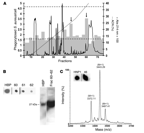
Figure 4. Identification of HBP and HNP1–3 as enhancers of bacterial phagocytosis in macrophages.
(A) Fractionation of PMN-sec by reversed-phase HPLC. PMN-sec was loaded onto a C18 column. Proteins (right y axis, solid curve) were eluted with a gradient of ACN with 0.1% TFA (right y axis, dotted line). Gray bars indicate the phagocytic activity resulting from stimulation with material of 3 consecutive fractions (left y axis). Basal phagocytosis and phagocytosis in response to PMN-sec are indicated by the lower and upper dashed line, respectively. Arrows indicate active fractions (45–47 and 60–62). For each analysis, 4 independent experiments were performed. (B) Immunological detection of HBP in fraction 60–62 using dot blot (left panel). These fractions were pooled and further analyzed with Western blot analysis (right panel). As positive control for both Western and dot blot, 40 ng recombinant HBP was used. (C) HPLC fractions were screened for the presence of HNP1–3 by dot blot analysis. Positive staining was detected in fractions 46 (insert). Inserts of dot blots in B and C were run on the same gel but were noncontiguous. As positive control 20 ng HNP1 was used. Determination of the mass values of material in fraction 46 with MALDI-MS gave molecular weights close to the theoretical values of HNP1–3.

