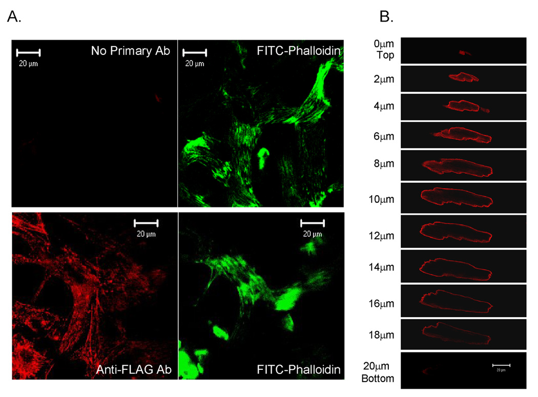
Figure 1. Immunolocalization of CRNK in neonatal and adult cardiomyocytes.

In Panel A, NRVM grown on Permanox® chamberslides were infected with Adv-CRNK (5moi, 24h). Cells were then fixed, permeabilized, stained with anti-FLAG monoclonal antibody (1:10) followed by rhodamine-conjugated goat anti-mouse secondary antibody, and counterstained with FITC-conjugated phalloidin to detect actin filaments. Control chamberslides were handled in an identical fashion, except that the anti-FLAG antibody was omitted (No Primary Ab). Cells were then viewed with a confocal microscope. In Panel B, ARVM were infected (100moi, 24h) with Adv-CRNK. Cells were then fixed, permeabilized and stained with anti-FLAG antibody followed by rhodamine-conjugated goat-antimouse secondary antibody. Cells were then optically sectioned (1µm) using the confocal microscope.
