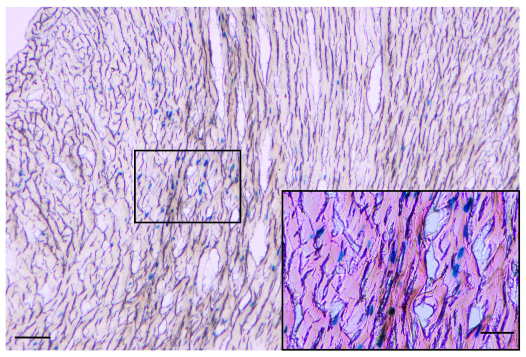
Figure 5. Adenoviral gene transfer of nuclear-encoded β-galactosidase into normal hearts.

Adv-neβgal (~1010 pfu) was injected by an Optison-mediated gene transfer technique into the aortic root of anesthetized rats in order to define the regions of myocardium transduced by the procedure. Following gluteraldehyde perfusion of whole hearts 3−4 days after gene transfer, tissue sections (14µm thick) were processed to detect β-galactosidase activity by X-gal staining. As is evident from this section of the LV posterior wall, scattered regions of nuclear β-galactosidase expression was detected throughout the full thickness of the myocardium. A high power image (box and insert) indicated that both cardiomyocytes and vascular endothelial cells expressed the transgene. Bar = 25µm.
