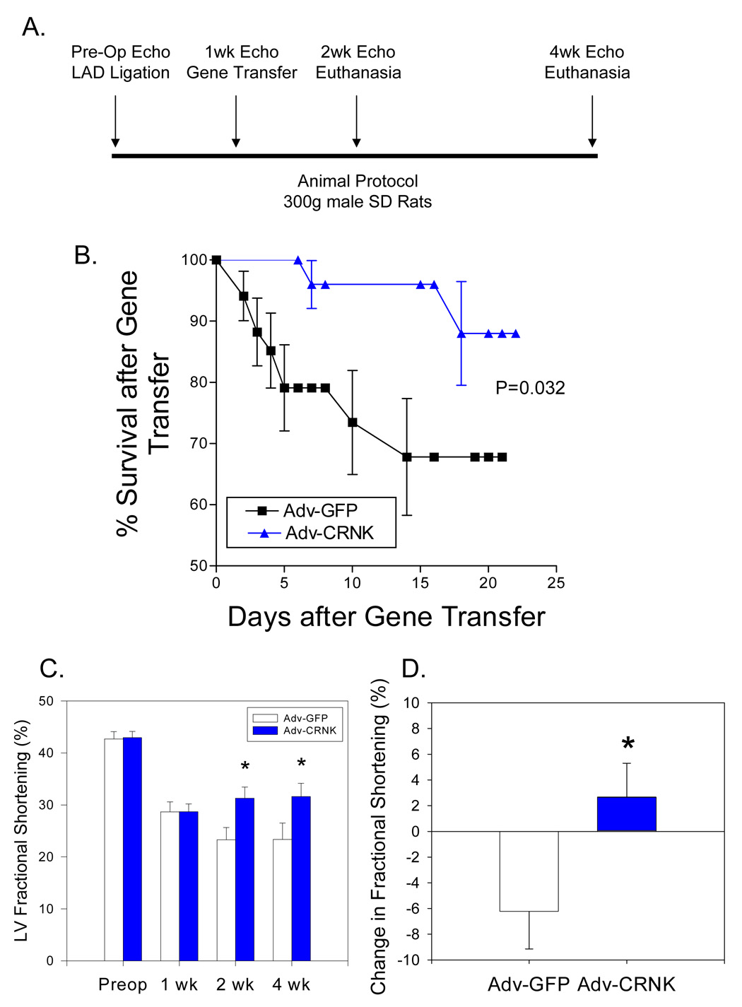
Figure 9. Adenoviral gene transfer of Adv-GFP and Adv-CRNK after myocardial infarction.

In Panel A, the gene transfer protocol is schematically outlined. Rats were subjected to coronary artery ligation and gene transfer with ~1010 pfu of either Adv-GFP (n=34) or Adv-CRNK (n=28). Surviving animals were then subjected to general anesthesia, echocardiography, and euthanasia at either 2 or 4 wks following infarct surgery (i.e., 1 or 3 wks after gene transfer). In Panel B, survival curves for Adv-GFP and Adv-CRNK animals were generated by the method of Kaplan and Meier, and compared using the log-rank test (P=0.032 for Adv-GFP vs. Adv-CRNK). In Panel C, LV fractional shortening measurements were compared in Adv-GFP vs. Adv-CRNK infected rats each time point in the gene transfer protocol. In Panel D, the change in fractional shortening between the 1wk and 2wk echocardiograms of Adv-GFP and Adv-CRNK infected rats are depicted. *P<0.05 vs. Adv-GFP at each time point examined.
