
Figure 4.
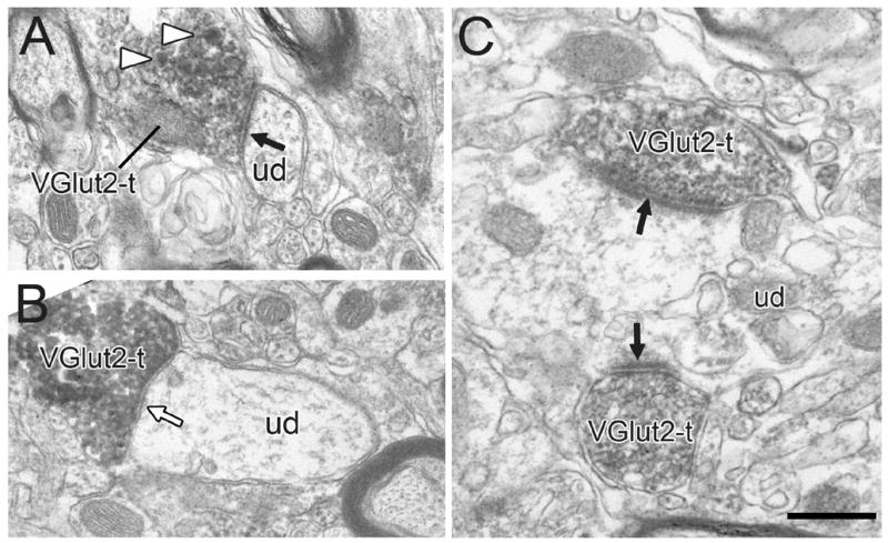
Electron micrographs showing typical VGlut2-labeled axon terminals (VGlut2-t). Most VGlut2-ts form asymmetric synapses (black arrows) onto unlabeled dendrites (ud), although others occasionally form symmetric synapses (white arrow). The VGlut2-t in panel A contains dense-cored vesicles (white arrowheads). Panel C shows an example of two VGlut2-ts forming convergent synapses onto a common dendrite. Scale bar, 0.5 μm in A, B; 0.4 μm in C.