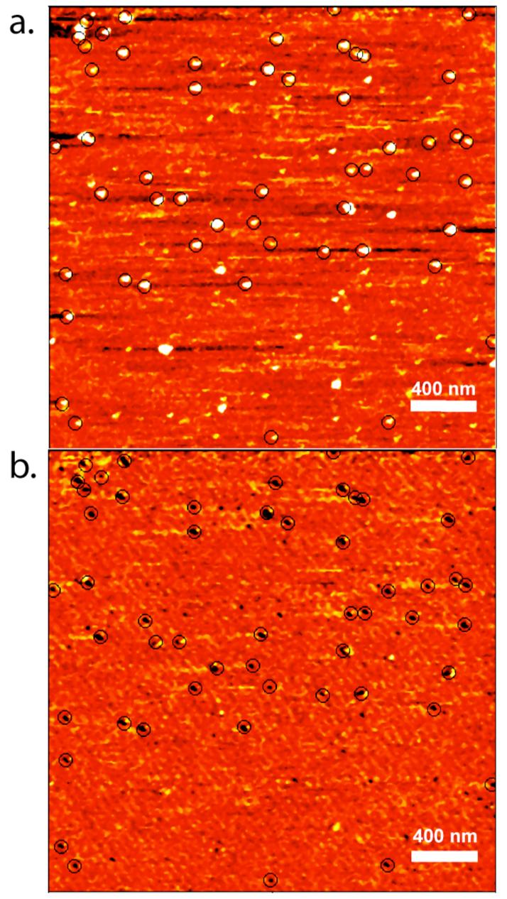
Figure 2.

Demonstration of aptamer recognition in recognition imaging microscopy. Topography (a) and recognition images (b) were simultaneously acquired for pure histone H4 protein deposited on a mica surface. Recognition events are shown with circles. In this image-pair, 53 out of 62 histone molecules are recognized by the anti-H4 aptamer 4.13.
