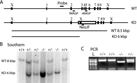
FIGURE 1.
Generation of MAGP-1 deficient mice. A, schematic of the Mfap2 locus in wild-type (WT) and MAGP-1-/- (KO) animals demonstrating mRNA disruption and genotyping strategies. Numbered rectangles correspond to Mfap2 exons 1–9, the open rectangle corresponds to the inserted neomycin resistance cassette. Short horizontal lines indicate the location of the Southern blot probe and PCR primers, boxes marked with “X” indicate XbaI restriction sites. Long horizontal lines represent fragments hybridizing to the Southern blot probe, 8.5 and 6 kbp for wild-type (WT) and MAGP-1-/- (KO), respectively. All distances, except the length of PCR primers, are to scale. B, XbaI-digested genomic DNA samples from a MAGP-1+/- cross were separated by agarose gel electrophoresis and detected by Southern blot. Resulting genotypes are indicated above each lane. C, genomic DNA was submitted to PCR with genotyping primers and separated by agarose gel electrophoresis. The predicted band sizes are 1160 and 714 bp for wild-type and MAGP-1-/-, respectively. L, 100-bp ladder.

