FIGURE 8.
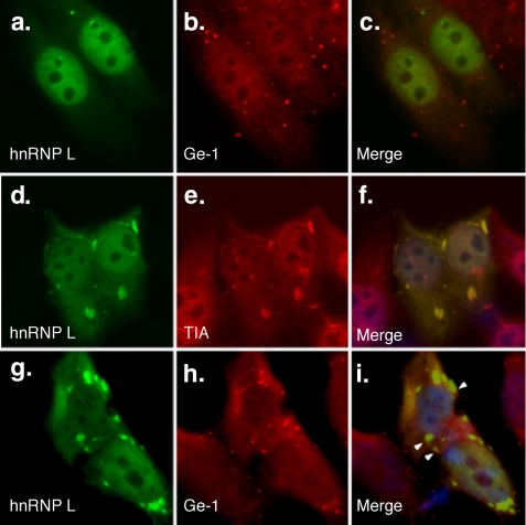
Trafficking of hnRNP L to stress granules but not processing bodies. After transfection into Hep-2 cells, green fluorescent protein-labeled hnRNP L localized to the nucleus (green; a). Serum containing antibodies directed against mRNA processing body marker Ge-1 stained 5-10 dots in the cytoplasm of Hep-2 cells (red; b). After exposure to arsenite, hnRNP L localized to granules in the cytoplasm (d) and co-localized with T-cell intracellular antigen (TIA), a marker for stress granules (red; e). mRNA-processing bodies (h) localized adjacent to hnRNP L in stress granules (g). Overlap of green and red staining is shown in c, f, and i. 4′,6-Diamidino-2-phenylindole staining in f and i indicate the location of cell nuclei. White arrowheads (i) indicate the location of Ge-1-containing mRNA processing bodies adjacent to hnRNP L-containing stress granules.

