
FIGURE 4.
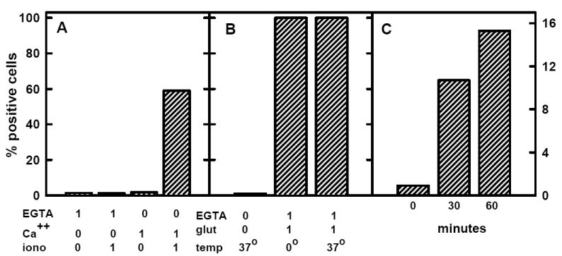
Annexin V binding to red cells. Panel A: Red cells were washed in HBS, suspended to 0.2 μl/ml (~2 × 10⁶ cells/ml) in HBS containing 0 or 1 mM EGTA, 0 or 1 mM CaCl2 and 0 or 1 μM ionomycin and incubated 30 min at 37 °C. The cells then were washed and analyzed for the binding of FITC-annexin V. Panel B: As in panel A, except that 0 or 1% w/v glutaraldehyde was the agent and the 30 min incubation was performed at 0 or 37 °C. Panel C: Red cells were washed and 3 aliquots suspended to 0.2 μl/ml (~2 × 10⁶ cells/ml) in 310 mM sucrose-5 mM histidine (pH 7.5). (This iso-osmotic buffer does lyse the cells or cause the leakage of hemoglobin.) The suspensions were incubated at 37 °C for the times indicated prior to chilling, washing and analysis. [Note that the full scale for panels A and B is 100% and for panel C is 16%.]