
Figure 8.
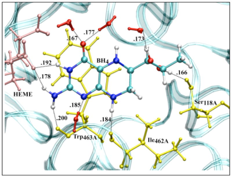
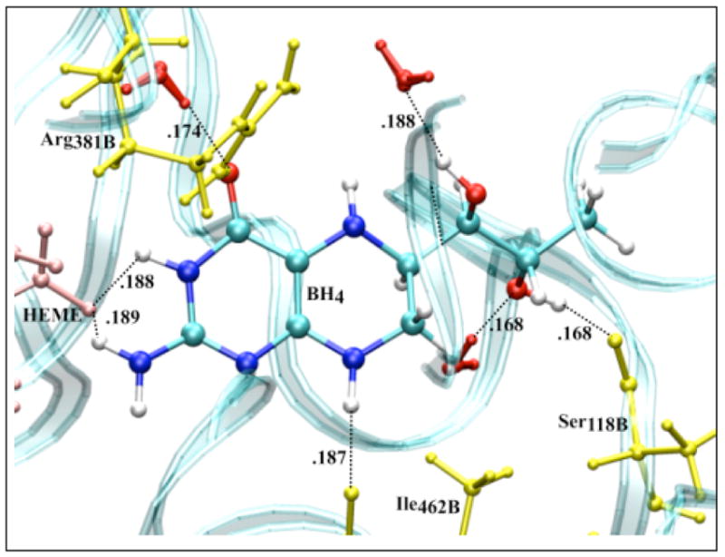
Amino acid residues (yellow), P450 heme (pink) and solvent (red) configuration around neutral BH4 bound to chain A (top) and chain B (bottom) of iNOSoxy dimer. The water (TIP4P) box containing a iNOSoxy dimer (OPLS) was energy minimized using Gromacs program. There are nine H-bonds (black dashed lines) between BH4 (chain A) and amino acid residues, heme and water within 0.2 nm, and seven H-bonds for BH4 bound to chain B.