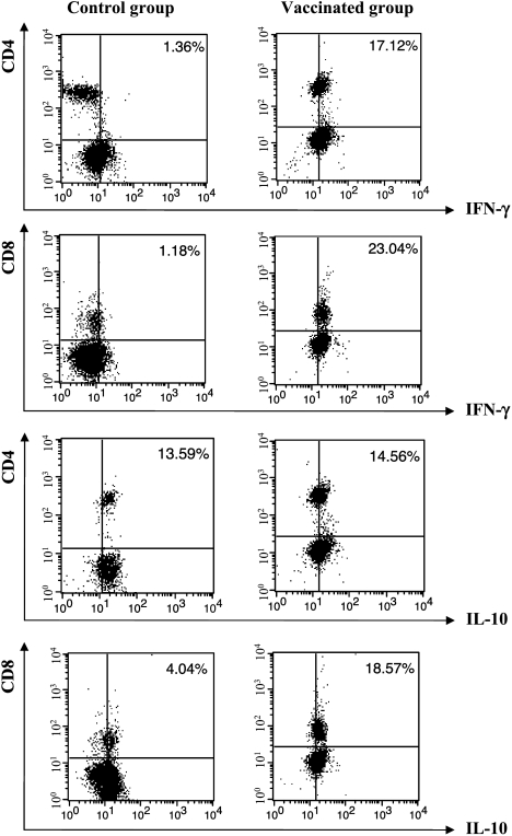
Figure 6.
Analysis of T lymphocyte population using FACS from tumor-bearing animals untreated (control group) and vaccinated with both plasmids in association with drug 7A. Cells were labeled with PE-conjugated anti-CD4 mAb or PE-conjugated anti-CD8 mAb. After permeabilization, intracellular detection of the cytokines was carried out with anti-cytokine biotinylated antibody and labeling with FITCconjugated streptavidin. Data are shown as percentages of double-positive cells (CD4/CD8 and IFN-γ/IL-10) and are representative of three independent experiments with similar results.

