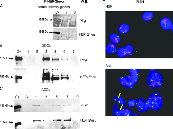
Figure 2.
HER-2/neu expression and activation. For each sample, protein lysate derived from the unbound proteins of anti-TRK-A IP was immunoprecipitated with α-HER-2/neu antibody, run on gel, and blotted with α-PTyr antibody (PTyr panel) for receptor phosphorylation status and with α-HER-2/neu antibody (HER-2/neu panel) for receptor expression. Lane C+: positive control (SKBR3 cell line). Lane numbers correspond to the cases reported in Table 1. (A) Expression of HER-2/neu and its phosphorylation in the two normal salivary glands that express the receptor. (B) Expression of HER-2/neu and its phosphorylation in six SDC cases. (C) Expression of HER-2/neu and its phosphorylation in six ACC cases. (D) HER-2/neu gene amplification analysis in SDC cases. Fluorescent in situ hybridization on imprint specimens. Chromosome 17 probe is labeled in Spectrum Green; HER-2/neu locus-specific probe is labeled in Spectrum Orange. Nuclei are counterstained with DAPI. Upper panel: amplification in HSR in SDC case No. 2 of Table 1. Lower panel: amplification in DM in SDC case No. 5 of Table 1. Single-copy gene signals are indicated by arrows.

