Figure 3.
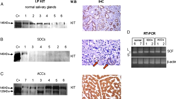
KIT expression and activation. For each sample, protein lysate derived from the unbound proteins of anti-TRK-A and anti-HER-2/neu IPs was immunoprecipitated with α-KIT antibody, run on gel, and blotted with α-KIT antibody (KIT panel) for receptor expression. Lane C+: positive control (Δ559 cell line). Lane numbers correspond to the cases reported in Table 1. (A) Expression of KIT in six normal salivary glands. KIT expression detected in these samples was in keeping with the immunohistochemistry decoration we observed (right panel), using Dako CD117 antibody. (B) Expression of KIT in six SDC cases. Absence of KIT expression by immunohistochemistry experiments was also detected in tumoral specimens; normal salivary duct and mastocytes, positive in-built controls, are indicated by arrows. (C) Expression of KIT in six ACC cases. The high KIT expression is confirmed by the strong CD117 positivity detected in these tumoral samples. (D) Expression of SCF transcript detected by RT-PCR experiment in normal and tumoral salivary glands. A total of 1 µl of cDNA was used as template for each SCF and β-actin gene amplification reaction; 10 µl of PCR were loaded on 2% agarose gel. In the case of SCF amplification, two bands of the expected molecular weight were detected (L = 494 bp and S = 409 bp).

