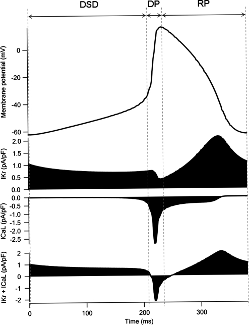
Fig. 5.
Simulated action potential and ionic currents of SA node cells. Simulated action potential and changes in IKr current, ICaL current, and sum of IKr and ICaL accompanying the spontaneous action potential are indicated. BCL of the action potential was 382 ms, which was shorter than that of early embryonic ventricular cells (492 ms). The MDP was approximately the same in the SA node cells (−62.17 mV) and early embryonic ventricular cells (−62.86 mV). The overshoot was 16.14 mV in the SA node cells: this value was more positive than that in the early embryonic ventricular cells (3.13 mV)

