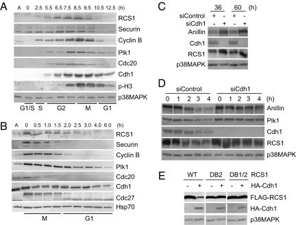
Fig. 3.
RCS1 is degraded in a Cdh1-dependent manner in vivo. (A and B) HeLa S3 cells were synchronized at the G1/S boundary by a double-thymidine arrest (A) or at prometaphase by a thymidine-nocodazole arrest (B). Cells were then released into fresh media and harvested at the indicated times. Levels of RCS1, securin, cyclin B, Plk1, Cdc20, Cdh1, Cdc27, phospho-histone H3 (Ser-10) (p-H3), p38MAPK (a loading control), and Hsp70 (a loading control) were analyzed by Western blotting. A, asynchronous cells. (C) HeLa cells were control-transfected or transfected with an siRNA against Cdh1 and harvested at the indicated times after transfection. Levels of anillin, Cdh1, RCS1, and p38MAPK were determined by Western blot analysis of total cell lysates. (D) HeLa cells were control-transfected or transfected with an siRNA against Cdh1 and arrested at prometaphase by incubating with nocodazole for 20 h. Cells were then released into fresh media in the presence of 100 μg/ml cycloheximide and harvested at the indicated times after release. Levels of anillin, Plk1, Cdh1, RCS1, and p38MAPK were determined by Western blot analysis. (E) FLAG-RCS1, FLAG-RCS1-DB2, or FLAG-RCS1-DB1/2 were transfected into HeLa cells with HA or HA-Cdh1. Levels of FLAG-RCS1, HA-Cdh1, and p38MAKP were determined by Western blot analysis.

